


Emergency
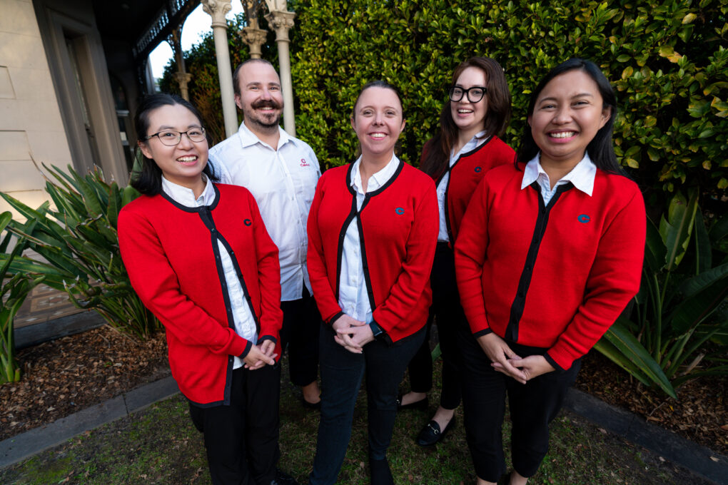
About us
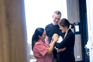
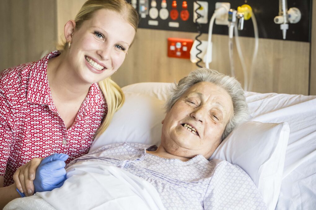
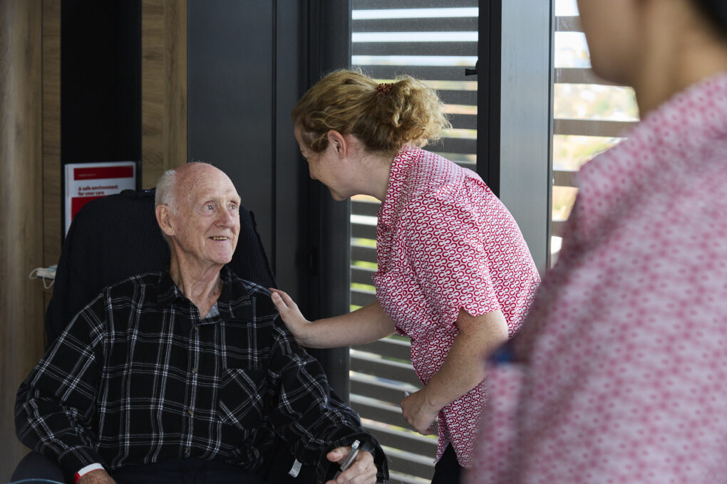
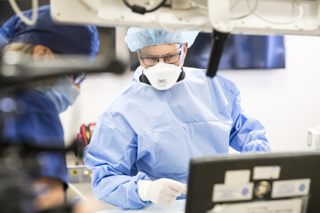
Cabrini Health is a Catholic, not-for-profit private health service located in Melbourne’s south east. Inspired by the mission and ethic of care of the Cabrini Sisters, we have been providing quality, compassionate care to our community for more than 70 years.
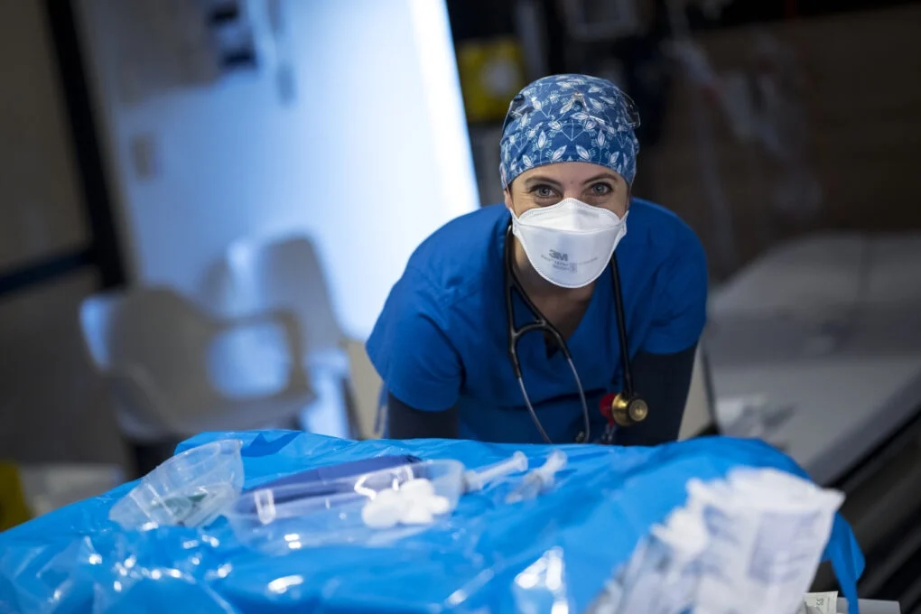
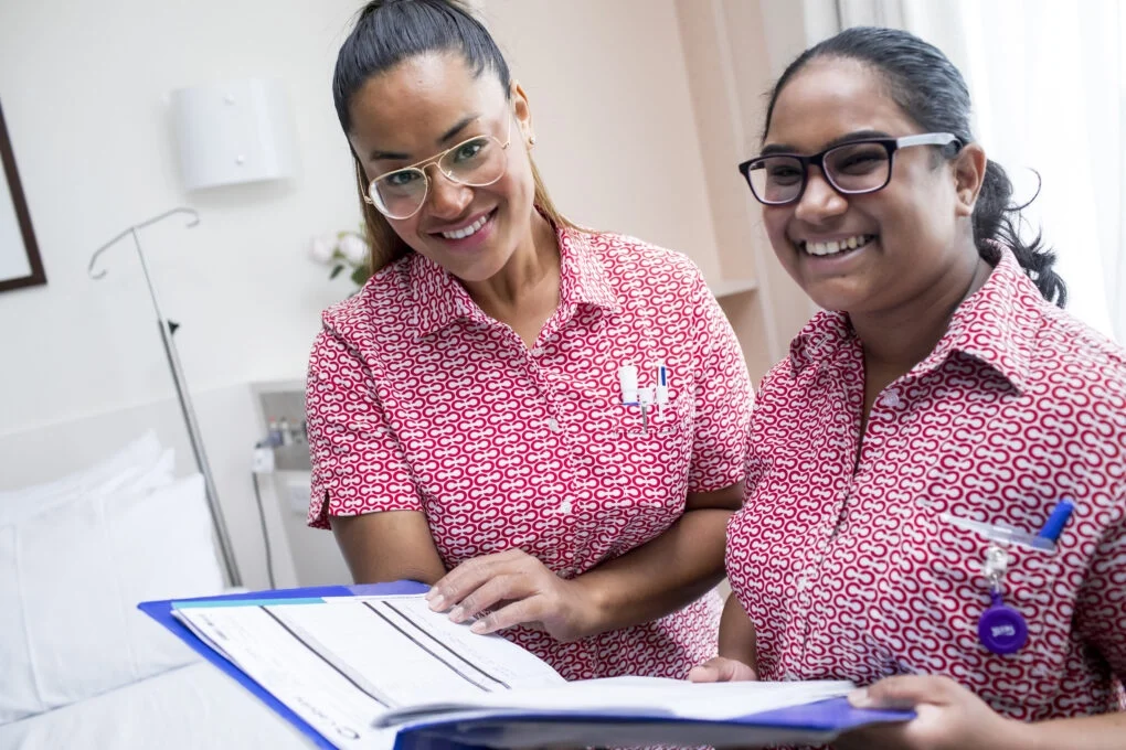
With hospitals in Malvern, Brighton and Elsternwick, Cabrini offers a comprehensive range of acute, rehabilitation, palliative care, mental health and homecare services.
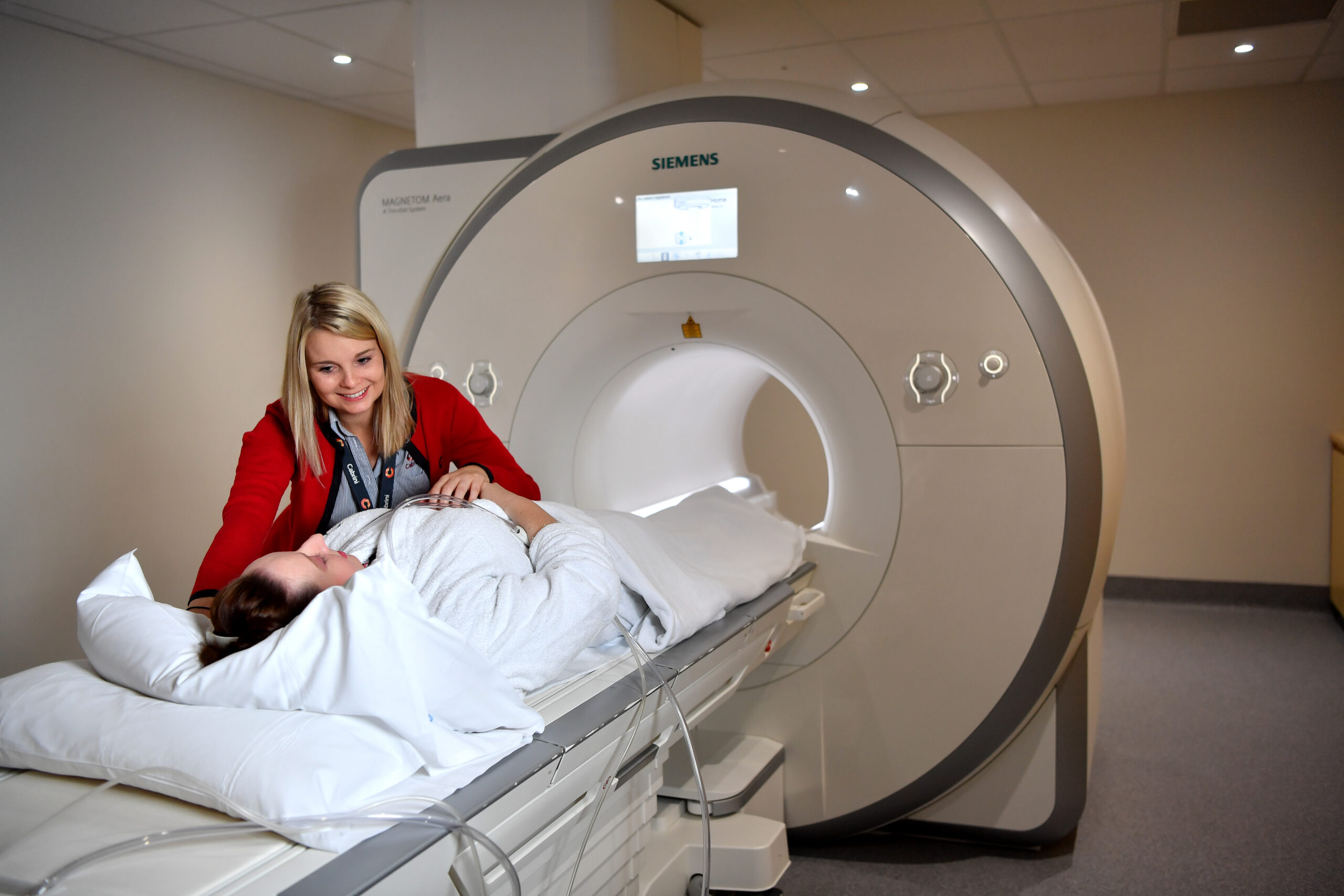
Our highly-skilled staff and specialists incorporate clinical research, innovative models of care and state-of-the-art technology to deliver the best possible care and outcomes for our patients.
Latest News